Cell Structure: notes and practice questions
- This topic covers cell theory, the structure of prokaryotic and eukaryotic cells, and microscopy techniques.
- Cells are the basic structural unit of all living organisms.
- Prokaryotic cells possess a cell wall, plasma membrane, cytoplasm, naked DNA in a loop, and 70S ribosomes.
- Eukaryotic cells feature a plasma membrane, compartmentalized cytoplasm with 80S ribosomes, a nucleus, and membrane-bound organelles such as mitochondria and endoplasmic reticulum.
- Microscopy skills include calculating actual size and magnification, and drawing annotated diagrams from micrographs.
- Unicellular organisms carry out all life processes: homeostasis, metabolism, nutrition, movement, excretion, growth, and reproduction.
How it is examined
One of the most reliably examined topics. Paper 1A tests identification and the prokaryote/eukaryote contrast. Paper 1B has used micrograph scale bars for calculate the actual size or calculate the magnification items. Paper 2 asks draw, label, annotate and distinguish, and drawing marks are lost for unruled label lines, labels that touch the wrong structure, and annotations with no function stated.
- A2.2.1 Cells as the basic structural unit of all living organisms. NOS: deductive reason generates predictions from theories, so cell theory predicts a newly discovered organism will consist of one or more cells.
- A2.2.2 Microscopy skills. Application of skills: temporary mounts, staining, measuring with an eyepiece graticule, coarse and fine focus, calculating actual size and magnification, producing a scale bar, taking photographs.
- A2.2.3 Developments in microscopy: advantages of electron microscopy, freeze fracture, cryogenic electron microscopy, fluorescent stains and immunofluorescence in light microscopy.
- A2.2.4 Structures common to cells in all living organisms: DNA as genetic material, cytoplasm mostly water, a plasma membrane of lipids. Students should understand the reasons for these.
- A2.2.12 Origin of eukaryotic cells by endosymbiosis. All eukaryotes evolved from a common unicellular ancestor that had a nucleus and reproduced sexually. Mitochondria evolved by endosymbiosis, and in some eukaryotes chloroplasts did too. Evidence: 70S ribosomes, naked circular DNA and the ability to replicate. NOS: a theory's strength comes from what it explains and predicts.
- A2.2.13 Cell differentiation as the process for developing specialized tissues in multicellular organisms, based on different patterns of gene expression often triggered by environmental change.
- A2.2.14 Evolution of multicellularity, which has evolved repeatedly. Many fungi and eukaryotic algae and all plants and animals are multicellular. The advantages are larger body size and cell specialization.
Guiding questions
- What are the features common to all cells and the features that differ?
- How is microscopy used to investigate cell structure?
Linking questions
- What explains the use of certain molecular building blocks in all living cells?
- What are the features of a compelling theory?
Practice questions
13 questions · 2 easy · 10 medium · 1 hardQuestion 1
EasyPaper 1A · calculator1 markWhich structure is found in prokaryotic cells but not in eukaryotic cells?
A. Plasma membrane
B. Cell wall
C. Nucleoid
D. Cytoplasm
Consider the defining features of prokaryotes. Which of these structures is exclusively found in them, and which are also present in at least some eukaryotes?
Question 2
MediumPaper 2 · calculator7 marksThe diagram shows part of a DNA molecule undergoing replication.
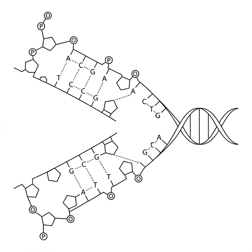
Identify two different types of chemical bonds shown in the DNA molecule.
(b) Explain the process of semi-conservative replication of DNA.
(c) Distinguish between the chromosomal DNA of prokaryotes and eukaryotes.
Look at the bonds that hold the two strands together, and the bonds that form the backbone of each individual strand.
Describe the key steps starting from the unwinding of the DNA double helix. Mention the roles of key enzymes and the principle of complementary base pairing. What does 'semi-conservative' mean in terms of the final product?
Think about the shape of the main chromosome, its location within the cell, and whether it is associated with any proteins.
Question 3
HardPaper 2 · calculator15 marksEukaryotic cells are characterized by the presence of membrane-bound organelles, which compartmentalize various biochemical processes.
(a) Compare and contrast the structure of chloroplasts and mitochondria.
(b) Describe the advantages of compartmentalization in eukaryotic cells.
(c) Explain the role of compartmentalization in the synthesis, modification, and transport of proteins destined for secretion.
Think about the membranes, genetic material, and internal structures of both organelles. Remember to provide both similarities and differences.
Consider how having separate 'rooms' in a cell might help with efficiency, optimal conditions, and protecting the cell from its own enzymes.
Trace the pathway of a protein from the DNA code in the nucleus to its release outside the cell. Mention the specific organelles and vesicles involved.
Question 4
EasyPaper 1A · calculator1 markWhich of the following is a characteristic of prokaryotic cells?
A. Their DNA is associated with histone proteins.
B. They have a compartmentalized cytoplasm.
C. They contain naked DNA in a loop.
D. They possess 80S ribosomes.
Recall the differences in genetic material, compartmentalization, and ribosome size between prokaryotes and eukaryotes.
Question 5
MediumPaper 2 · calculator4 marksThe diagram shows a simplified prokaryotic cell and a eukaryotic animal cell.
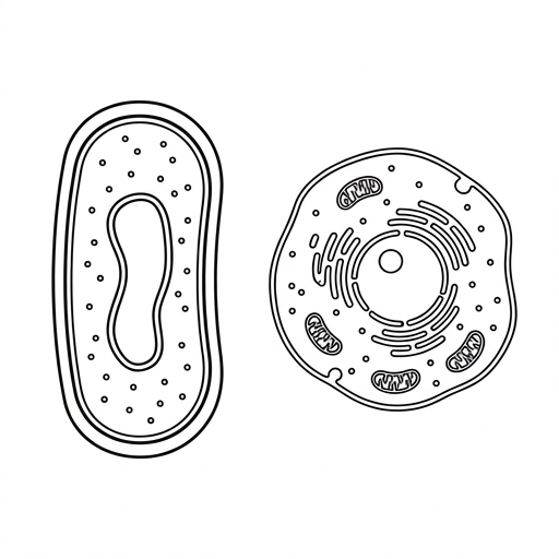
(a) State two structures that are present in both the prokaryotic cell and the eukaryotic animal cell.
(b) (i) State one membrane-bound organelle present in the eukaryotic cell but absent in the prokaryotic cell.
(b) (ii) State one difference, other than location, between the DNA of the prokaryotic cell and the DNA of the eukaryotic cell.
Look for components that are fundamental to any cell's basic function and boundary, regardless of its complexity.
Think about the key feature that defines a eukaryotic cell, which is the presence of internal compartments.
Consider the shape of the DNA molecule and what it is associated with inside the cell.
Question 6
MediumPaper 1A · calculator1 markStriated muscle fibres are considered an exception to some principles of the cell theory. What is a key feature of these cells that makes them atypical?
A. They have a much larger volume than typical eukaryotic cells.
B. They contain multiple nuclei enclosed within a single plasma membrane.
C. They lack the ability to undergo mitosis for repair.
D. They are composed of repeating contractile units called sarcomeres.
Consider the basic principles of the cell theory, such as 'all living things are composed of cells' and what a 'cell' is typically defined as. Which feature of a muscle fibre does not fit this typical definition?
Question 7
MediumPaper 1A · calculator1 markA micrograph of a bacterium is shown. The image of the bacterium is 50 mm long. The scale bar in the image is 20 mm long and represents an actual distance of 2 µm.
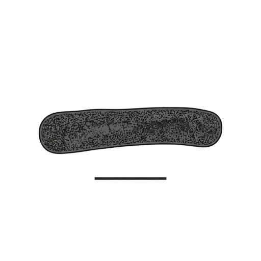
What is the magnification of the micrograph?
A. ×1 000
B. ×10 000
C. ×25 000
D. ×50 000
Remember the formula for magnification: Magnification = Image size / Actual size. Be careful to convert all measurements to the same unit before calculating. Use the information given for the scale bar to find the magnification, not the length of the bacterium.
Question 8
MediumPaper 1B · calculator6 marksA student is studying a cross-section of a leaf from a sunflower (Helianthus annuus) using a light microscope. They focus on a single palisade mesophyll cell. The actual length of this cell is 30 µm. The student draws the cell with a length of 6 cm in their notebook.
(a) Calculate the magnification of the student's drawing.
(b) Distinguish between the magnification and the resolution of a microscopic image.
(c) The student wants to observe the detailed internal structure of a chloroplast from the palisade cell, including the thylakoid stacks. State the type of microscope required for this task.
(d) State one reason for the high density of chloroplasts in palisade mesophyll cells.
Remember the formula for magnification: Magnification = Image size / Actual size. Make sure your units for image size and actual size are the same before you divide.
Think about what each term describes. One refers to the size of the image, and the other refers to its clarity or detail. A good answer will define both and highlight the difference.
Consider the limitations of a light microscope. What kind of microscope can see much smaller structures in high detail?
What is the main function of a palisade cell in a leaf, and which organelle is responsible for this function?
Question 9
MediumPaper 2 · calculator5 marksThe image shows a transmission electron micrograph (TEM) of a chloroplast.
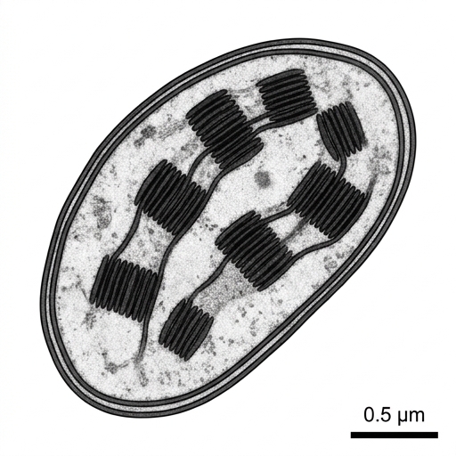
(a)
(i) Name a type of cell in a flowering plant that does not contain chloroplasts.
(a) (ii) State two products of the overall process of photosynthesis.
(b) Chloroplasts contain their own circular DNA and 70S ribosomes. Explain how these features provide evidence for the endosymbiotic theory.
Consider the different parts of a plant. Which parts are not exposed to sunlight and therefore have no need for photosynthesis?
Recall the word equation for photosynthesis. The products are the substances that are created during the reaction.
Compare the features mentioned (circular DNA, 70S ribosomes) with the features of prokaryotic and eukaryotic cells. What does the term 'endosymbiosis' imply about the origin of this organelle?
Question 10
MediumPaper 1A · calculator1 markA scientist is studying three microorganisms (P, Q, and R) isolated from a soil sample. The presence (+) or absence (–) of several cellular structures was recorded for each, as shown in the table.
| Feature | Organism P | Organism Q | Organism R |
|---|---|---|---|
| Nucleus | – | + | + |
| 80S ribosomes | – | + | + |
| Mitochondria | – | + | + |
| Chloroplasts | – | + | – |
| Cell wall | + | + | – |
Based on the data, what are the most likely classifications for these microorganisms?
| P | Q | R | |
|---|---|---|---|
| A. | Prokaryote | Plant | Animal |
| B. | Fungus | Plant | Animal |
| C. | Prokaryote | Animal | Plant |
| D. | Plant | Prokaryote | Animal |
First, use the presence or absence of a nucleus to distinguish between prokaryotes and eukaryotes. Then, use the other organelles, such as the cell wall and chloroplasts, to differentiate between the types of eukaryotes.
Question 11
MediumPaper 1A · calculator1 markMature human red blood cells and mature phloem sieve tube elements both lack a nucleus.
What is a direct consequence of this atypical structure?
A. They cannot divide by mitosis
B. Their genetic material is free in the cytoplasm
C. They are unable to produce ATP
D. They lack a plasma membrane
Think about what a nucleus contains and what cellular processes rely directly on the presence of a nucleus.
Question 12
MediumPaper 1A · calculator1 markThe table shows the presence or absence of certain features in three types of cells (X, Y and Z).
| Feature | Cell X | Cell Y | Cell Z |
|---|---|---|---|
| Mitochondria | present | absent | present |
| Cell wall | present | present | absent |
| 80S ribosomes | present | absent | present |
| Chloroplasts | present | absent | absent |
Which row correctly identifies the types of cells?
| Cell X | Cell Y | Cell Z | |
|---|---|---|---|
| A. | Plant | Prokaryote | Animal |
| B. | Plant | Animal | Prokaryote |
| C. | Prokaryote | Plant | Animal |
| D. | Animal | Prokaryote | Plant |
Consider which cell types possess membrane-bound organelles like mitochondria and chloroplasts, and recall the difference in ribosome size between prokaryotes and eukaryotes.
Question 13
MediumPaper 1A · calculator1 markResearchers recently determined the precise three-dimensional structure of the spike protein from a novel virus. To achieve this high-resolution image, they rapidly cooled the protein sample to to prevent the formation of damaging water crystals, keeping the protein molecules stable in their natural conformation.
Which technique did the researchers use?
A. Cryogenic electron microscopy
B. Freeze-fracture electron microscopy
C. X-ray diffraction
D. Immunofluorescence
Consider which technique involves freezing a sample to preserve the natural state of individual molecules for high-resolution imaging, rather than splitting a membrane or using antibodies.
No question on this page matches those filters. Try another difficulty or paper.
Every Cell Structure question, marked for you
Every answer is marked mark by mark, IB-style, and the AI tutor helps when you are stuck.
Where marks are lost
- Command terms are consistently under-read. Students answer describe when the question said explain, so they give an account with no reasons and cap at half marks. The reverse also happens on outline, where a student writes an essay for a 2-mark summary and runs out of time.
- compare and contrast answers give only similarities, or only differences.